Breast Cancer Diagnosis

“I feel a breast lump. What next?”

“Is it cancer?”

“What is breast biopsy?”

First appointment with the doctor

If you notice a lump or any change in your breast, you will need to have an assessment to identify what has caused the change.

So, when a lady arrives at our clinic, with painless breast lump or any change in breasts, first of all, we relieve her anxiety and tell her not to worry. Then, we carry out a comprehensive triple assessment to confirm whether lump is cancer or non-cancerous. This robust assessment will relieve your anxiety and give you peace of mind.

In Triple-Assessment…

(1) You will be assessed by Dr Rajinder Kaur Saggu, (Female Breast Specialist doctor), who will assess your risk factors by taking a detailed clinical history and performing an examination of both breasts and armpit.

(2) A breast imaging doctor will do Ultrasound and/or Mammogram. (*Please note that women over age of 40 may require a Mammogram).

(3) If an abnormality is identified in either of these scans, needle aspiration or a core biopsy will be performed.

triple assessment test
“Are there any additional tests I need?”

If an abnormality in your breast is identified, you will likely require further tests. This can range from Image-Guided Needle Biopsy, MRI and/or other Imaging Investigations.

A range of genetic tests and Counselling are also available, and we are happy to discuss the options with you.

We are happy if you are accompanied by a partner, friend of relative to your appointment.

What is Breast Biopsy?

A breast biopsy is a procedure to remove a sample of breast cells or tissue, or an entire lump.

A pathologist then looks at the sample under the microscope to check further signs of the disease.

A biopsy is the only way to find out if cells are cancerous or not.

Biopsy is usually done in an office or a clinic or an OPD basis. This means you will go home the same day as the procedure. Local anesthesia is used in some biopsies. This means you will be awake, but you won’t feel pain in your breast during the procedures.

Try not to Panic or worry

If breast cancer is found on biopsy, it can be treated. With standard treatment, most people with breast cancer found early have a good prognosis (high chance of survival).

Types of Breast Biopsy

(1) Fine needle aspiration (FNA)

This is a simple procedure that takes only few minutes. Your doctor will insert a thin needle into the breast to take out fluid and cells.

The problem with FNA is if malignant cells are seen, the pathologist may not be ale to tell if the cancer is invasive or non invasive. So if cancer is diagnosed on FNA, additional biopsies may be necessary.

(2) Core needle biopsy

Also called Core Biopsy, it uses a needle to remove cylindrical pieces or cores of breast tissue. The samples are about the size of a grain of a rice.

Core biopsy is an OPD based procedure.

• It is done under local anesthesia.
• A small injection is given over the area of lump to make it numb, and then, a special hollow needle is inserted into the lump and few cores of the tissue are taken out.
• The cores are sent to a pathologist for biopsy to confirm the diagnosis.
• A core biopsy not only confirms the cancer, but also tells us which ‘type’ it is.
• We routinely assess 3 IHC markers for each and every breast cancer patient on biopsy samples- ER (Estrogen Receptor), PR (Progesterone Receptor) and HER2neu.
• The information obtained helps us to plan the further treatment.

If mammogram is used to guide the breast biopsy it’s called a stereotactic biopsy. Otherwise it is called an ultrasound-guided core needle biopsy or MRI-guided core needle biopsy. The procedure is usually done by a radiologist but can also be performed by a surgeon.

“Relax, core needle breast biopsies procedures are common and most patients tolerate them very well.”

(3) Excisional Biopsy

When an entire lump or suspicious area is removed, it is called Excisional or Open Surgical Biopsy.

Breast lesions that cannot be felt or palpated but are only seen on imaging need wire-localization. A very thin guide-wire is passed through the skin into area of concern. After localization the patient is taken to the operating. An incision is made in the skin, the wire is followed to the mass or calcifications and the tissue is removed. Most of the time, the specimen is X-rayed to be sure the correct spot was removed.

Then the tissue is sent to the pathologist for “biopsy”.

(4) Incisional Biopsy

If the breast mass is large, only a part of the mass can be removed for diagnosis. This is an open incisional biopsy.

(5) Vacuum Assisted Breast Biopsy (VABB)

In this procedure a hollow probe is put through a small cut into the abnormal area of breast tissue using an image guidance.

A core of tissue is then suctioned into the probe & a rotating knife inside the probe cuts the tissue samples.

This method removes more tissue than a regular core needle biopsy.

What is Biopsy Report?

The biopsy report comes within 2-5 working days, I assess it in detail, sit with the patient and the family, and explain to them line by line.

If it reveals cancer, Patient and family is informed about the diagnosis and I assure them that “Breast cancer treatment is far more advanced as compared to before. We are together in this and I shall guide you and walk you through each step of this journey”.

Also try not to panic or worry, as with standard treatment, early breast cancers are curable.

I discuss all cases in a Multi-Disciplinary Meeting and develop a personalised treatment plan specific to you.

What is Staging Test?

Once breast cancer diagnosis is confirmed, we wish to know how much it has spread in the body. The lesser it has spread, the earlier is the stage. And earlier stages have a better outcome than later stage.

Tests and procedures used to stage breast cancer may include:

  • Ultrasound abdomen
  • Chest X ray
  • Breast MRI
  • Bone scan
  • Computerized tomography (CT) scan
  • Positron emission tomography (PET) scan


Not all women will need all of these tests and procedures. We will select the appropriate tests based on your specific circumstances.

Stages of Breast Cancer

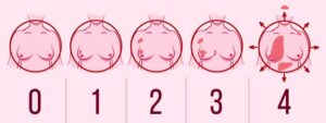
Once the diagnosis of cancer is established by radiology and pathology, then staging is done to determine its extent. Staging describes a cancer based on how much cancer is there in the body and where it is when first diagnosed. Different treatment strategies can be used depending on the stage of breast cancer.

Stages can be determined as :

• STAGE 0 : DCIS (ductal carcinoma in situ). Cancer cells are trapped inside breast ducts with no ability to spread. LCIS ( lobular carcinoma in situ) is not cancer, despite the name.

• STAGE IA/IB : Cancer cells invade the walls of duct or lobule, but the total size is under 2 cm, cells have not spread to lymph nodes.

• STAGE IIA/IIB : Cancers over 2 cm that have not spread to nodes or invaded chest muscle; Cancers under 5 cm in size that have spread to 1 to 3 axillary (armpit) lymph nodes.

• STAGE IIIA/IIIB : Cancers of any size that spread to 4 or more axillary (armpit) lymph nodes, the nodes around the clavicle (collarbone), and/or the nodes under the sternum (internal mammary nodes); cancers over 5 cm that spread to any nodes; and tumours that have grown into the chest wall.

• STAGE IV : Cancer has spread beyond the breast and nearby nodes to other organs or distant nodes. The most commonly involved sites are lung, liver, brain and bones.